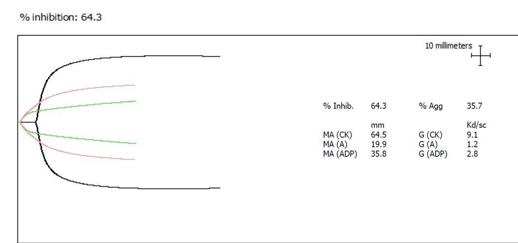
Here are some follow-up comments to Dr. Larry Brace’s August 11, 2016 post about monitoring Plavix reversal with platelet concentrate.
From Robert Gosselin, SSCLS, UC Davis: The thromboelastograph is not sensitive to platelet dysfunction. We could consider using their Platelet Mapping (PLTMAP) method, but there is no reference or therapeutic range provided, and the only thing the package insert provides is a linearity type study. The ADP agonist concentration is low at 5uM. Our reference range for ADP agonist mapping is <60% and for arachidonic acid agonist is <20%. The method is too sensitive to AA/P2Y12 inhibitors, but may be useful in the particular case, although I am sure there are no outcome studies looking at PLTMAP test for P2Y12 reversal using PLT transfusions.
And from Oksana Volod, MD, Cedars-Sinai, Los Angeles: In my lab I have TEG PLTMAP, whole blood aggregometry, and the Accriva VerifyNow. Based on my personal experience with over 5000 TEG PLTMAP interpretations, I do rely a lot on TEG platelet mapping. It shows not only the percent of platelet inhibition but also the overall picture. I do not rely on percent inhibition, however, I rely instead on the MA-A and MA-ADP values. Thus, MA-ADP >50 mm I would consider hemostatic. The patient whose pattern is shown below is at risk of bleeding during surgery with an MA-ADP of 35.8 mm. You have to pay attention to the MA-A value. In our hands if it is over 40 mm, you can’t interpret TEG PLTMAP. TEG alone was never designed to assess platelet function.
Mr. Gosselin asked, “What is your MA reference range for P1, P2 and P3?”
Dr Volod replied,
P1 is the activator (as labelled on vial) = MA-A. This is contribution of fibrinogen to the clot. Per Haemonetics it should not exceed 25 mm for a valid interpretation. Per our study comparing whole blood aggregometry and TEG PM I am comfortable to report MA-AA and MA-ADP values if MA-A is <40 mm. The basic TEG MA reference range is 50–70 mm. MA-AA and MA-ADP represents residual platelet activity . So if they are >50 mm, I consider this hemostatic and if <50 mm, adequately anticoagulated.
P2 is ADP (as labelled on vial) = MAAA
P3 is AA (as labelled on vial) = MADP
Dr. Volod also provided this reference, click or tap for complete article: Mahal E, Suarez TA, Bliden KP, et al. Platelet function measurement–based strategy to reduce bleeding and waiting time in clopidogrel-treated patients undergoing coronary artery bypass graft surgery; the timing based on platelet function strategy to reduce clopidogrel-associated bleeding related to CABG (TARGET-CABG) study. Circulation Cardiovascular Interventions 2012; 5: 261–9.
A follow-up from Vadim Kostousov, PhD: TEG platelet mapping assay might be compromised in the presence of heparin (click or tap):
Nelles NJ, Chandler WL. Platelet mapping assay interference due to platelet activation in heparinized samples. Am J Clin Pathol 2014;142:331–8.
Dr. Volod responded, “This is why I pointed MA-A cut off value :)”


No comments here.